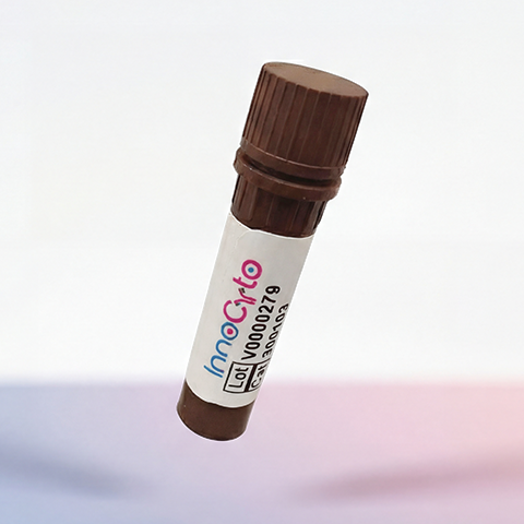
| Cat # | Size | Price | Quantity | |
|---|---|---|---|---|
| 633401 | 100 μg | $300 | ||
| 633402 | 500 μg | $900 |
| Application | Bioassay |
|---|---|
| Format | Lyophilized from sterile PBS, pH7.4. |
| Expression Host | HEK293 |
| Target Name | FceR1a, FceR1-a, high affinity IgE receptor, FCER1A, FceR1 alpha,High affinity immunoglobulin epsilon receptor subunit alpha |
| Species | Human |
| accession number | P12319 |
| Sources | A DNA sequence encoding the human FCER1A (P12319) (Met1-Gln205) was expressed with a C-terminus His tag. |
| Molecular Weight | The protein has a predicted MW of 47.8 KDa. Due to glycosylation, the protein migrates to 70-78 KDa based on Bis-Tris PAGE result. |
| Affinity Tag | C-His |
| Purity | ≥95% as determined by SDS-PAGE. ≥90% as determined by SEC-HPLC |
| Regulatory Status | RUO |
| Endotoxin level | < 1.0 EU per μg protein |
| Protein Concentration | Lyophilized |
| Storage and Handling | Samples are stable for up to twelve months from date of receipt at -20℃ to -80℃. Store it under sterile conditions at -20℃ to -80℃. It is recommended that the protein be aliquoted for optimal storage. Avoid repeated freeze-thaw cycles. |
Human Fc epsilon RI alpha (FcεRIα), also known as FCER1A or the high-affinity IgE receptor alpha subunit, is a transmembrane glycoprotein that plays a central role in allergic immune responses. It is expressed primarily on mast cells and basophils, where it serves as the ligand-binding component of the high-affinity receptor for immunoglobulin E (IgE). FcεRIα helps initiate immune responses against allergens and contributes to the rapid release of inflammatory mediators.
FcεRIα is a type I transmembrane protein belonging to the immunoglobulin superfamily. Its extracellular region contains two immunoglobulin-like domains that form the high-affinity IgE-binding site, followed by a single transmembrane segment and a short cytoplasmic region. On mast cells and basophils, FcεRI typically forms a tetrameric complex consisting of one α subunit, one β subunit, and two disulfide-linked γ subunits. The α subunit binds IgE, while the β and γ subunits contribute to receptor expression and intracellular signal transduction.
The principal ligand of FcεRIα is the Fc region of IgE. When allergen-specific IgE binds to FcεRIα on the cell surface, subsequent allergen exposure can cross-link adjacent IgE–receptor complexes. This activates intracellular signaling pathways, leading to mast-cell and basophil degranulation and the release of histamine, leukotrienes, prostaglandins, and inflammatory cytokines. These mediators contribute to immediate hypersensitivity reactions and allergic inflammation.
Dysregulated FcεRI signaling is associated with allergic diseases, including allergic asthma, allergic rhinitis, atopic dermatitis, food allergies, and anaphylaxis. Because FcεRIα is essential for IgE-mediated activation, it represents a potential target for therapeutic intervention. Strategies under investigation include blocking IgE–FcεRI interactions, inhibiting receptor activation, and developing soluble FcεRIα proteins to sequester IgE. These approaches may help reduce allergic inflammation and provide new opportunities for treating IgE-mediated disorders.
Recombinant Human Fc epsilon RI alpha/FCER1a Protein (c-His) TDS
Have a product or application question? Consult our FAQs or contact us.