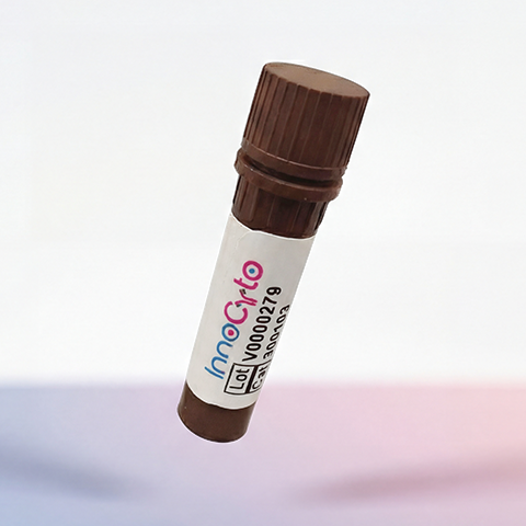
| Cat # | Size | Price | Quantity | |
|---|---|---|---|---|
| 670901 | 10 μg | $175 | ||
| 670902 | 50 μg | $340 |
| Application | Bioassay |
|---|---|
| Format | Lyophilized from sterile PBS, pH 7.4. |
| Expression Host | HEK293 |
| Target Name | IL-3, Interleukin-3, Burst promoting activity, Eosinophil colony stimulating factor (Eo-CSF) |
| Species | Mouse |
| accession number | NP_034686.2 |
| Sources | A DNA sequence encoding the mouse IL3 (NP_034686.2) (Met1-Cys166) was expressed. |
| Molecular Weight | The recombinant mouse IL3 consists of 140 amino acids and predicts a molecular mass of 15.7 KDa. The molecular weight of this protein is around 19.5 KDa verified by SEC-MALS (Routinely tested). |
| Affinity Tag | None |
| Purity | > 90 % as determined by SDS-PAGE. |
| Regulatory Status | RUO |
| Endotoxin level | < 0.1 EU per μg protein |
| Protein Concentration | Lyophilized |
| Storage and Handling | Samples are stable for up to twelve months from date of receipt at -20℃ to -80℃. Store it under sterile conditions at -20℃ to -80℃. It is recommended that the protein be aliquoted for optimal storage. Avoid repeated freeze-thaw cycles. |
Mouse interleukin-3 (IL-3) is a hematopoietic cytokine that regulates the proliferation, survival, differentiation, and activation of blood-forming cells. It is produced primarily by activated T lymphocytes and mast cells in response to immune stimulation. IL-3 supports the development of multiple hematopoietic lineages, including granulocytes, monocytes, basophils, eosinophils, and mast cells, and helps coordinate immune and inflammatory responses.
Mouse IL-3 is a secreted glycoprotein belonging to the class I hematopoietic cytokine family. Its mature form contains approximately 140 amino acids and adopts a characteristic four-helix bundle structure. The protein contains two intramolecular disulfide bonds and potential N-linked glycosylation sites, which may influence its biochemical properties. As a soluble cytokine, IL-3 binds to specific cell-surface receptors to initiate intracellular signaling rather than functioning as a membrane-bound ligand.
The mouse IL-3 receptor consists of an IL-3-specific alpha subunit (IL-3Rα, CD123) and a signaling beta subunit. Unlike humans, mice express two beta subunits: the common beta chain (βc, CD131), which is shared with the receptors for granulocyte-macrophage colony-stimulating factor (GM-CSF) and interleukin-5 (IL-5), and an IL-3-specific beta chain (βIL-3). The latter can bind mouse IL-3 directly, although productive signaling requires the receptor alpha subunit. Receptor activation stimulates pathways such as JAK2/STAT5, PI3K/AKT, and MAPK, promoting cellular growth, survival, and functional activation.
Dysregulated IL-3 signaling contributes to allergic inflammation, asthma, immune-mediated disorders, and experimental models of inflammatory disease. IL-3 may also support the growth and survival of certain leukemic cells, making its receptor system relevant to hematologic cancer research. Therapeutically, IL-3 signaling is being investigated as a target for controlling excessive inflammation, while recombinant IL-3 has been studied for its ability to stimulate hematopoietic recovery after bone marrow suppression. In preclinical research, mouse IL-3 is valuable for studying cytokine biology, hematopoiesis, immune regulation, and potential cytokine-targeted therapies.
Recombinant Mouse IL-3 Protein TDS
Have a product or application question? Consult our FAQs or contact us.