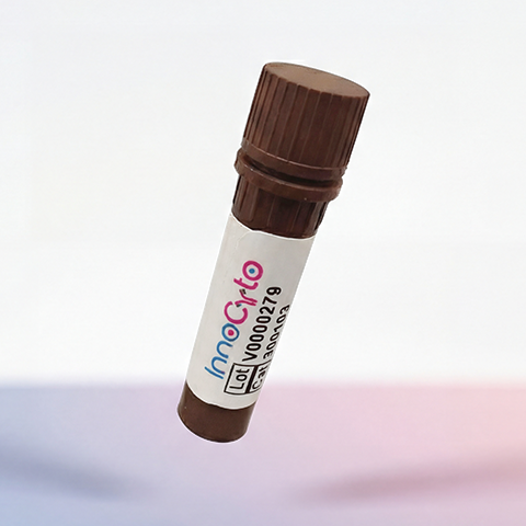
| Cat # | Size | Price | Quantity | |
|---|---|---|---|---|
| 670801 | 100 μg | $540 | ||
| 670802 | 1 mg | $2500 |
| Application | Bioassay |
|---|---|
| Format | Lyophilized from sterile 50 mM Tris, 300 mM NaCl, 1 mM DTT, pH8.0 |
| Expression Host | E.coli |
| Target Name | IL-33, IL-1F11 |
| Species | Mouse |
| accession number | Q8BVZ5 |
| Sources | A DNA sequence encoding the Mouse IL33 (Q8BVZ5) (Ser109-Ile266) was expressed, with a polyhistidine tag at the N-terminus. |
| Molecular Weight | The recombinant Mouse IL33 consisting of 165 amino acids and has a calculated molecular mass of 18.53 KDa. |
| Affinity Tag | None |
| Purity | ≥95% as determined by SDS-PAGE. |
| Regulatory Status | RUO |
| Endotoxin level | < 0.1 EU per μg protein |
| Protein Concentration | Lyophilized |
| Storage and Handling | Samples are stable for up to twelve months from date of receipt at -20℃ to -80℃. Store it under sterile conditions at -20℃ to -80℃. It is recommended that the protein be aliquoted for optimal storage. Avoid repeated freeze-thaw cycles. |
Mouse interleukin-33 (IL-33) is a member of the interleukin-1 (IL-1) cytokine family that functions as both an immune signaling molecule and an alarmin released in response to cellular stress or tissue damage. It is constitutively expressed in various structural cells, including epithelial and endothelial cells and fibroblasts, and can be released following tissue injury. IL-33 promotes type 2 immune responses by activating group 2 innate lymphoid cells (ILC2s), T-helper 2 (Th2) cells, mast cells, eosinophils, and other immune populations. Its activities include stimulating cytokine production, supporting tissue repair, and regulating inflammatory responses.
Mouse IL-33 is encoded by the Il33 gene and is synthesized as a protein containing an N-terminal nuclear localization region and a C-terminal IL-1-like cytokine domain. Unlike many secreted cytokines, full-length IL-33 can localize to the nucleus, where it may associate with chromatin. When released extracellularly, IL-33 binds to its receptor ST2 (also called IL1RL1), which associates with the IL-1 receptor accessory protein (IL-1RAcP) to form a functional signaling complex. Receptor activation triggers pathways involving MyD88, IRAKs, and NF-κB and MAP kinases. Soluble ST2 acts as a decoy receptor that can bind IL-33 and limit its signaling.
IL-33 has important roles in allergic inflammation, asthma, atopic dermatitis, and other type 2 inflammatory conditions. Elevated or dysregulated IL-33 signaling can promote eosinophilic inflammation, mucus production, and airway hyperresponsiveness. It also participates in responses to parasitic infections and can support tissue repair and immune regulation. Depending on the tissue and disease context, IL-33 may have beneficial or harmful effects.
Therapeutically, IL-33 and ST2 are targets of interest for treating asthma and other inflammatory diseases. Blocking IL-33 or its receptor may reduce excessive type 2 immune responses, while controlled activation of this pathway is being studied for tissue repair and selected immune applications. Mouse IL-33 is widely used to investigate cytokine signaling, allergic disease, innate immunity, and potential immunomodulatory therapies.
Recombinant Mouse IL-33 Protein TDS
Have a product or application question? Consult our FAQs or contact us.