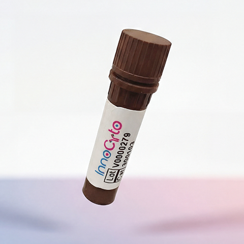
| Cat # | Size | Price | Quantity | |
|---|---|---|---|---|
| 802103 | 25 ug | $245 | ||
| 802104 | 100 ug | $595 |
| Application | Flow Cytometry |
|---|---|
| Format | Liquid, APC |
| Expression Host | CHO |
| Target Name | GPC3, GTR2-2, MXR7, SDYS, OCI5, |
| Species | Human |
| Sources | Human Glypican-3 (Gln25-His559) with C-terminus His tag is expressed in CHO cells and conjugated to APC. |
| Accession Number | P51654 |
| Molecular Weight | The protein has a predicted molecular weight of 62kDa. Under DTT-reducing conditions, it migrates at approximately 65-100 kDa on SDS-PAGE prior to conjugation. |
| Affinity Tag | C-His |
| Regulatory Status | RUO |
| Formulation | 1xPBS buffer, pH7.4, 0.09% NaN3 with a carrier protein |
| Endotoxin level | Not tested |
| Protein Concentration | 25µg size is bottled at 0.1mg/mL concentration. 100 µg size is bottled at lot specific concentration. |
| Storage and Handling | Briefly centrifuge the vial upon receipt. An unopened vial may be stored at 2–8°C for up to six months. |
| Research Areas | Development, Embryogenesis, Cancer Marker |
Glypicans are heparan sulfate proteoglycans anchored to the cell surface by a glycosyl-phosphatidylinositol (GPI) linkage. There are six known mammalian glypicans (GPC1 to GPC6), which can be released by the lipase Notum. They regulate signaling pathways like Wnt, Hedgehog, FGF, and BMPs. Glypican-3 (GPC3) plays a role in modulating IGF2 interactions and influences cell proliferation. It also interacts with FGF-basic via heparan sulfate chains. Mutations in GPC3 cause Simpson-Golabi-Behmel Syndrome (SGBS), leading to overgrowth, dysmorphism, and malformations. GPC3 is expressed as a 70 kDa precursor, cleaved into a 40 kDa N-terminal and 30 kDa C-terminal subunit. It is highly expressed in Hepatocellular carcinoma and melanoma, but downregulated in cancers like ovarian, cholangiocarcinoma, mesothelioma, and breast cancer due to promoter hypermethylation.
APC Human Glypican-3 Protein (C-His) TDS
Human Glypican-3 Protein (C-His)
Biotin Human Glypican-3 Protein (C-His)
PE Human Glypican-3 Protein (C-His)
Have a product or application question? Consult our FAQs or contact us.