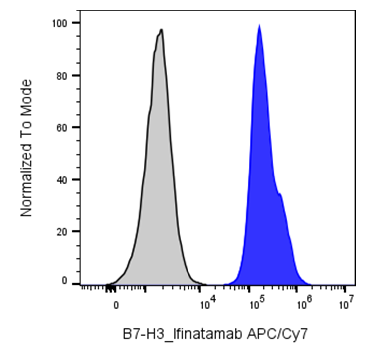
| Cat # | Size | Price | Quantity | |
|---|---|---|---|---|
| 113007 | 25 tests | $135 | ||
| 113008 | 100 tests | $295 |
| Clone | Ifinatamab |
|---|---|
| Application | Flow Cytometry |
| Reactivity | Human |
| Format | APC/Cyanine7 |
| Target Name | CD276, B7-H3, B7H3, B7RP-2 |
| Isotype | Human IgG1 |
| Antibody Type | Monoclonal |
| Regulatory Status | RUO |
| Formulation | Phosphate-buffered solution, pH 7.2, containing 0.09% sodium azide and 0.2% (w/v) BSA |
| Protein Concentration | Supplied at a lot-specific concentration. |
| Storage & Handling | The antibody solution should be stored undiluted between 2°C and 8°C, and protected from prolonged exposure to light. Do not freeze. |
| Recommended Usage | For flow cytometric staining, it is recommended to use 5 µL of this reagent per 0.5-1.0 million cells in a 100 µL volume. Optimal reagent performance should be determined by titration for each specific application. |
| Excitation Laser | Red Laser (633 nm) |
| See All Formats | Clone Ifinatamab |
B7-H3, also known as CD276, is a member of the B7 family of immune regulatory molecules that modulate T cell responses. Initially described as a co-stimulatory molecule that enhances T cell activation and interferon-γ production, subsequent studies have shown that B7-H3 can also exert inhibitory effects on immune responses. Its precise function appears to depend on the cellular context and microenvironment, with evidence supporting a predominantly immunosuppressive role in many settings.
Structurally, human B7-H3 is a type I transmembrane protein composed of extracellular immunoglobulin (Ig)-like domains, a transmembrane region, and a short cytoplasmic tail. In humans, the predominant isoform contains four Ig-like domains (4Ig-B7-H3), generated through exon duplication, whereas a two-domain isoform (2Ig-B7-H3) is more common in other species. The cytoplasmic domain lacks well-defined signaling motifs, suggesting that B7-H3 primarily signals through interactions with receptors on other cells.
The exact receptor (or receptors) for B7-H3 has not been definitively established, which remains a key area of investigation. Several candidates have been proposed, but none have been universally confirmed. Despite this, functional studies demonstrate that B7-H3 can regulate T cell proliferation, cytokine production, and cytotoxic activity, as well as influence natural killer (NK) cell function.
In disease, B7-H3 is highly expressed in a wide range of cancers, including prostate, lung, breast, and pediatric solid tumors, and its expression is often associated with poor prognosis, tumor progression, and immune evasion. It is also implicated in non-malignant conditions such as autoimmune and inflammatory diseases, although its role in these contexts is less well defined.
Therapeutically, B7-H3 is an attractive target in immuno-oncology. Approaches include monoclonal antibodies, antibody-drug conjugates, and chimeric antigen receptor (CAR) T cells designed to target B7-H3-expressing tumor cells. These strategies aim to overcome tumor immune evasion and enhance anti-tumor immunity, and several are currently under clinical investigation.
APC/Cyanine7 Human IgG1 isotype control
APC/Cyanine7 Anti-Human CD276 (B7-H3) Antibody TDS
Have a product or application question? Consult our FAQs or contact us.