| Cat # | Size | Price | Quantity | |
|---|---|---|---|---|
| 202103 | 25 tests | $80 | ||
| 202104 | 100 tests | $170 |
| Clone | IM7 |
|---|---|
| Application | Flow Cytometry |
| Reactivity | Mouse |
| Format | iF647 |
| Target Name | CD44, Epican, Extracellular matrix receptor III (ECMR-III), Phagocytic glycoprotein 1 (PGP-1) |
| Isotype | Rat IgG2b |
| Antibody Type | Monoclonal |
| Regulatory Status | RUO |
| Formulation | Phosphate-buffered solution, pH 7.2, containing 0.09% sodium azide and 0.2% (w/v) BSA |
| Protein Concentration | Supplied at a lot-specific concentration. |
| Storage&Handling | The antibody solution should be stored undiluted between 2°C and 8°C, and protected from prolonged exposure to light. Do not freeze. |
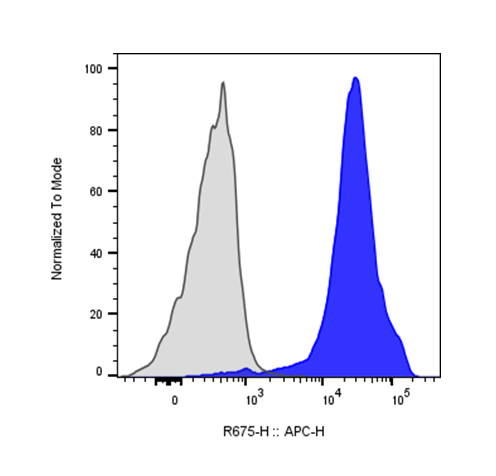
| Recommended Usage | For flow cytometric staining, it is recommended to use 5 µL of this reagent per 0.5-1.0 million cells in a 100 µL volume. Optimal reagent performance should be determined by titration for each specific application. iF647 has an excitation max at 656 nm and an emission max at 670 nm. |
| Excitation Laser | Red Laser (633 nm) |
| RRID | AB_3739077 |
| Research Areas | Adhesion Molecules, Cancer Marker, B cells, T cells, Monocytes, Macrophages, Astrocytes, Microglia |
| See All Formats | Clone IM7 |
CD44 is a widely expressed cell surface glycoprotein that functions as a receptor for extracellular matrix components and plays a central role in cell adhesion, migration, and signaling. It is expressed on many cell types, including leukocytes, epithelial cells, endothelial cells, and fibroblasts. In the immune system, CD44 is best known as an activation and memory marker on T cells, where it contributes to lymphocyte trafficking, tissue retention, and immune surveillance.
Structurally, CD44 is a type I transmembrane protein belonging to the hyaluronan receptor family. The extracellular domain contains a conserved N-terminal hyaluronan-binding domain, followed by a stem region that is subject to extensive alternative splicing. This alternative splicing generates multiple CD44 isoforms, commonly grouped into the standard form (CD44s) and variant isoforms (CD44v), which differ in ligand-binding capacity and tissue distribution. CD44 has a single transmembrane region and a cytoplasmic tail that interacts with cytoskeletal adaptor proteins such as ezrin, radixin, and moesin, linking CD44 to actin dynamics and intracellular signaling pathways.
The primary ligand for CD44 is hyaluronan, a major component of the extracellular matrix. CD44 can also bind other ligands, including osteopontin, collagens, fibronectin, matrix metalloproteinases, and certain growth factors. Ligand engagement regulates cell adhesion and motility and can activate signaling pathways involved in proliferation, survival, and inflammation. Through these interactions, CD44 helps coordinate immune cell recruitment to inflamed tissues and supports tissue remodeling and wound healing.
CD44 plays important roles in disease. In chronic inflammatory and autoimmune conditions, dysregulated CD44–hyaluronan interactions contribute to persistent immune cell infiltration and tissue damage. CD44 is also strongly implicated in cancer biology; many tumors upregulate CD44, particularly variant isoforms, which are associated with enhanced invasion, metastasis, and resistance to therapy. CD44 is widely used as a marker of cancer stem-like cells in multiple malignancies, including breast, colorectal, and hematologic cancers.
Therapeutically, CD44 is relevant as both a biomarker and a drug target. Antibodies and small molecules targeting CD44 or its interaction with hyaluronan have been explored to limit inflammation and tumor progression. CD44 is also being exploited for targeted drug delivery, as hyaluronan-based carriers can selectively accumulate in CD44-expressing tumors. In immunotherapy and immunomonitoring, CD44 expression is routinely used to identify activated and memory T cell populations, underscoring its broad importance in research and clinical applications.
iF647 Rat IgG2b Isotype Control Antibody
iF647 Anti-Mouse/Human CD44 Antibody TDS
iF488 Anti-Mouse/Human CD11b (Integrin αM) Antibody, Clone M1/70
FITC Anti-Mouse CD11c (integrin αX) Antibody, Clone N418
APC Anti-Mouse CD49b (pan-NK, integrin α2) Antibody, Clone DX5
iF560 Anti-Mouse CD11c (integrin αX) Antibody, Clone N418-mG1
Have a product or application question? Consult our FAQs or contact us.