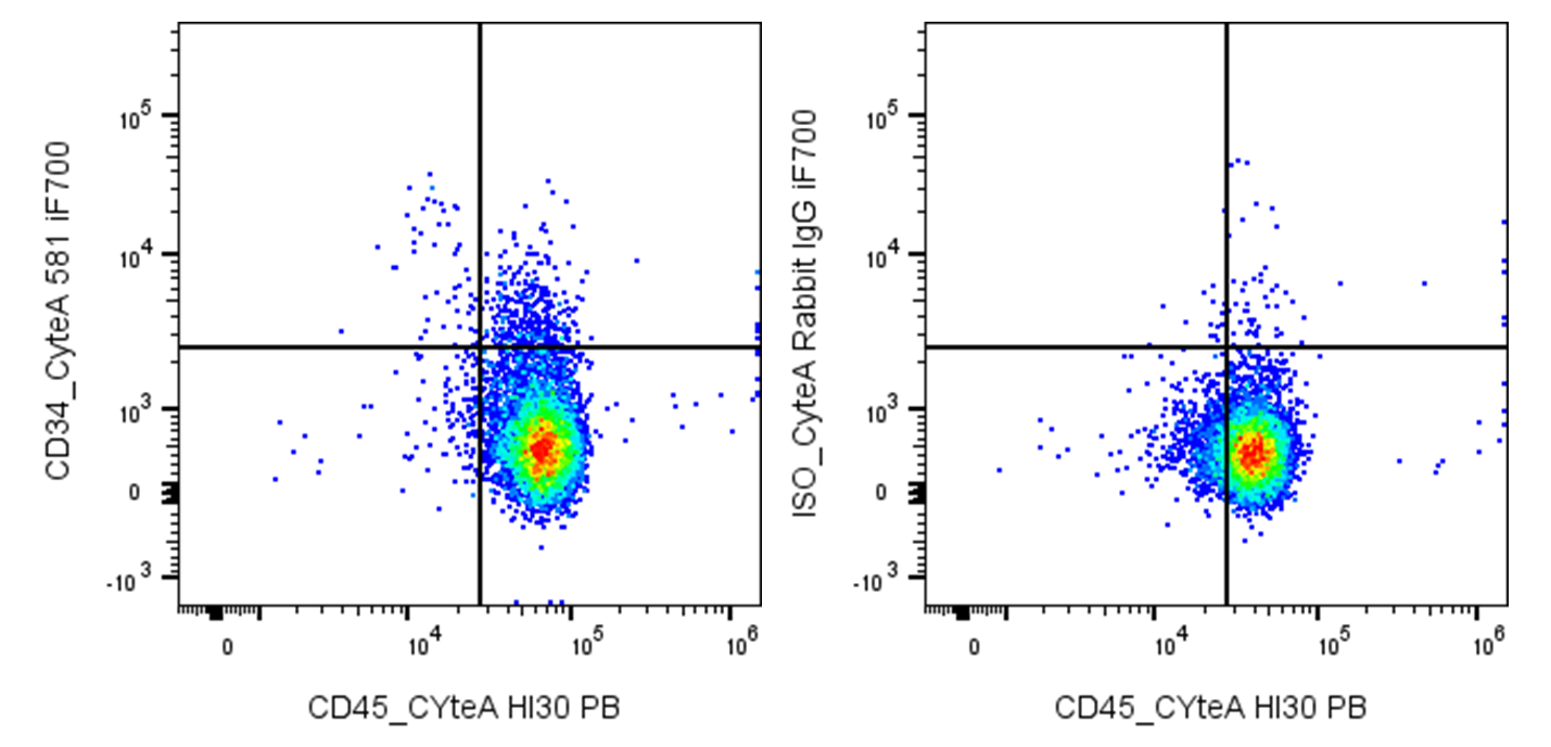
| Cat # | Size | Price | Quantity | |
|---|---|---|---|---|
| 180803 | 25 tests | $125 | ||
| 180804 | 100 tests | $250 |
| Clone | 034CB |
|---|---|
| Application | Flow Cytometry |
| Reactivity | Human |
| Format | iF700 |
| Target Name | CD34, Gp105-120, My10 |
| Isotype | Rabbit IgG |
| Antibody Type | Monoclonal |
| Regulatory Status | RUO |
| Formulation | Phosphate-buffered solution, pH 7.2, containing 0.09% sodium azide and 0.2% (w/v) BSA |
| Protein Concentration | Supplied at a lot-specific concentration. |
| Storage & Handling | The antibody solution should be stored undiluted between 2°C and 8°C, and protected from prolonged exposure to light. Do not freeze. |
| Recommended Usage | For flow cytometric staining, it is recommended to use 5 µL of this reagent per 0.5-1.0 million cells in a 100 µL volume. Optimal reagent performance should be determined by titration for each specific application. iF700 has an excitation max at 690 nm and an emission max at 710 nm. |
| Excitation Laser | Red Laser (633 nm) |
| Brand | InnoSite |
| See All Formats | Clone 034CB |
CD34 is a transmembrane phosphoglycoprotein that functions as a key marker of hematopoietic stem and progenitor cells, endothelial cells, and some fibroblast subtypes. It plays a vital role in cell adhesion, migration, and the maintenance of stem cell niches within the bone marrow. CD34 is widely used in clinical and research settings as a surface antigen for identifying and isolating hematopoietic stem cells, which are essential for bone marrow transplantation and regenerative therapies.
Structurally, CD34 is a single-pass transmembrane sialoglycoprotein of approximately 110 kDa, consisting of an extensively glycosylated extracellular domain, a single hydrophobic transmembrane segment, and a short cytoplasmic tail containing phosphorylation sites. The extracellular domain carries multiple O-linked and N-linked carbohydrate chains contributing to its highly negative charge, which is important for its anti-adhesive properties and interaction with other molecules. Although CD34 belongs to the sialomucin family, its precise molecular ligands have not been fully defined. It is known to interact with L-selectin and other adhesion molecules, particularly under vascular shear stress, facilitating leukocyte rolling and homing to lymphoid tissues or sites of inflammation.
In the context of disease, CD34 expression and function are significant in several pathological conditions. In hematologic malignancies such as acute myeloid leukemia (AML), CD34 is often expressed on leukemic stem cells, where it serves as a diagnostic and prognostic marker. In solid tumors, its expression is associated with angiogenesis, as CD34 marks endothelial progenitors contributing to new vessel formation. Furthermore, its downregulation or loss may relate to fibrotic progression in certain chronic disorders.
Therapeutically, CD34 has become a cornerstone in stem cell-based interventions. CD34-positive cells are collected and transplanted to regenerate healthy hematopoietic and immune systems following chemotherapy or radiation therapy in cancer patients. Clinical trials are also exploring the use of CD34+ progenitor cells to promote vascular repair and tissue regeneration in ischemic heart disease, peripheral artery disease, and diabetic ulcers. Efforts to modulate CD34 expression or exploit CD34+ cells’ regenerative capacity continue to shape the landscape of advanced cellular therapy and biotechnology.
InnoSite™ iF700 Rabbit IgG, κ2 Isotype Control
InnoSite™ iF700 Anti-human CD34 Antibody TDS
Have a product or application question? Consult our FAQs or contact us.